
Renal & Aortic Stenting
Renal Artery Stenting
🩺 What is Renal Artery Stenosis?
Renal Artery Stenosis is a condition where the arteries that carry blood to the kidneys become narrowed or blocked, usually due to fatty deposits (atherosclerosis). This reduces blood supply to the kidneys, leading to uncontrolled high blood pressure and kidney damage.
🔹 Signs and Symptoms
High blood pressure that is difficult to control with medicines
Worsening kidney function (high creatinine levels)
Sudden episodes of breathlessness due to fluid overload
Swelling in ankles and legs
In severe cases, recurrent episodes of flash pulmonary edema
🔹 Causes
Atherosclerosis (plaque buildup) – most common cause
Fibromuscular dysplasia – abnormal artery structure, often in younger patients
Rarely, vasculitis or trauma
🔹 Diagnosis
Doppler Ultrasound of kidneys to assess blood flow
CT Angiography / MR Angiography for detailed imaging
Renal Angiography – gold standard to confirm narrowing
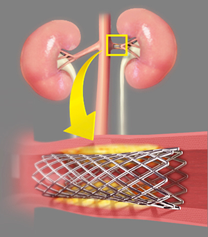
🔹 Treatment – Renal Artery Stenting
A catheter is guided to the narrowed portion of the artery.
A balloon is inflated to open the blockage.
A stent (metal mesh tube) is placed to keep the artery open.
🔹 Benefits
Better control of blood pressure
Protection of kidney function
Reduction in risk of heart failure due to hypertension
👉 Renal Artery Stenting is a safe and effective solution that restores blood flow to the kidneys and improves overall health.
🌟 Aortic Stenting (Endovascular Aneurysm Repair – EVAR)
🩺 What is Aortic Stenosis / Aneurysm?
The aorta is the largest artery in the body, carrying blood from the heart to the rest of the body. Sometimes, the aorta may develop:
Aortic Aneurysm – abnormal bulging or ballooning of the vessel wall.
Aortic Dissection – a tear in the inner wall of the aorta.
Both conditions can be life-threatening if not treated promptly.
🔹 Signs and Symptoms
Often silent in early stages
Back, abdominal, or chest pain
Sudden severe pain (tearing or ripping sensation) in cases of dissection
Dizziness, fainting, or shock in rupture cases
🔹 Causes
Long-standing hypertension
Atherosclerosis (cholesterol plaques)
Genetic conditions (Marfan’s, Ehlers-Danlos syndrome)
Trauma or infection
🔹 Diagnosis
Ultrasound / CT Angiography for detecting aneurysm size and location
MRI for detailed vascular imaging
Aortic Angiography for interventional planning
🔹 Treatment – Aortic Stenting (EVAR/TEVAR)
A catheter is inserted through the groin arteries.
A stent-graft (metal mesh with fabric) is placed inside the weakened aorta.
This reinforces the aortic wall, preventing rupture or further tearing.
🔹 Benefits
Minimally invasive alternative to open surgery
Reduced risk of rupture and life-threatening bleeding
Shorter hospital stay and quicker recovery
Effective long-term protection for patients with aneurysm or dissection
👉 Aortic Stenting is a life-saving procedure that provides durable results and improves patient survival in high-risk vascular conditions.
It can be somewhat more difficult to diagnose chronic kidney disease at an early stage because usually, the symptoms associated with condition do not become visible until after the condition has been in existence for quite some time. Anemia and hyperphosphatemia are some of the conditions which might accompany chronic kidney disease in the longer term and are considered symptoms for the same. There are a number of factors which experts consider to diagnose acute renal failure, including a sudden increase in creatinine and blood urea nitrogen levels.
A detailed medical evaluation is necessary for proper diagnosis of the condition and establishing if the condition can be classified as acute renal failure or chronic kidney disease. Ultrasound of kidneys might also be required to find out the nature of the condition and differentiate acute and chronic kidney disease. If the condition is diagnosed at an early stage, there are higher chances of arresting the growth of condition to decrease the possibility of the condition resulting in end-stage renal disease which can be difficult to deal with.