NASD / PDA Closure
Atrial Septal Defect (ASD) Closure
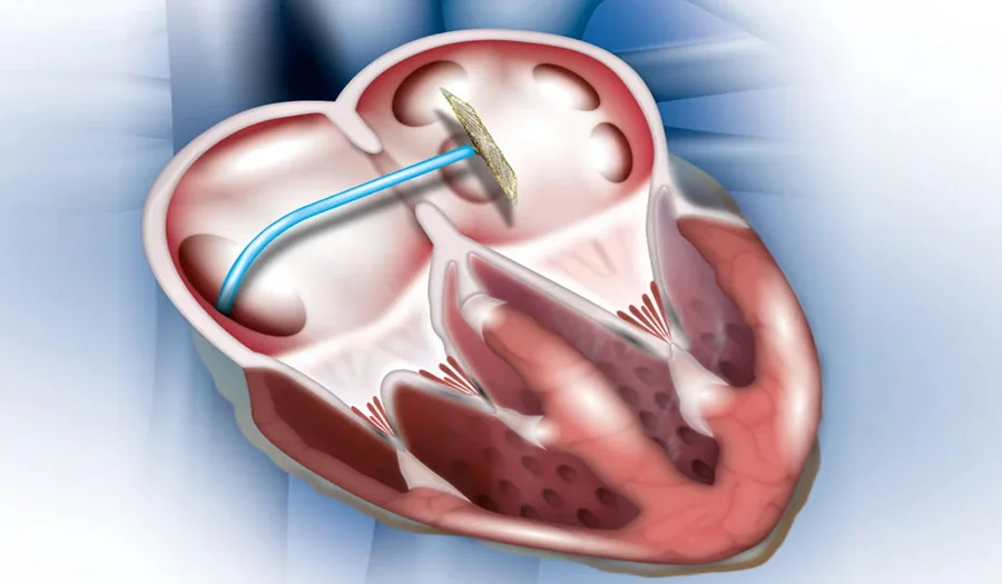
🫀 What is ASD?
An Atrial Septal Defect (ASD) is a hole in the wall (septum) between the two upper chambers of the heart (atria). This abnormal opening allows oxygen-rich blood to flow from the left atrium into the right atrium, mixing with oxygen-poor blood and putting extra strain on the heart and lungs.
🔹 Signs and Symptoms
Shortness of breath, especially during exercise
Fatigue and reduced stamina
Frequent lung infections
Heart murmurs (detected during clinical examination)
In severe or untreated cases: swelling in the legs, irregular heartbeats, or pulmonary hypertension
🔹 Causes
Congenital defect (present since birth) due to incomplete closure of the atrial septum
Sometimes associated with genetic conditions like Down’s syndrome
🔹 Diagnosis
Echocardiography (2D Echo/Doppler) is the gold standard for confirming ASD and measuring its size
ECG may show rhythm abnormalities
Chest X-ray can reveal enlarged heart chambers or lung congestion
🔹 Treatment – ASD Closure
Small ASDs may close on their own in early childhood.
For persistent or large ASDs, treatment is necessary to prevent complications.
Device closure: A minimally invasive procedure where a catheter is inserted via the groin to place a closure device in the defect.
Surgical closure: In cases where device closure is not possible, open-heart surgery may be performed.
👉 Successful ASD closure restores normal blood circulation, reduces heart strain, and allows patients to live a healthy, active life.
🌟 Patent Ductus Arteriosus (PDA) Closure
🫀 What is PDA?
The Ductus Arteriosus is a blood vessel that connects the pulmonary artery to the aorta in unborn babies, allowing blood to bypass the lungs. Normally, this vessel closes shortly after birth. When it remains open (patent), it is called Patent Ductus Arteriosus (PDA). This leads to abnormal blood flow between the aorta and pulmonary artery, overloading the heart and lungs.
🔹 Signs and Symptoms
Fast breathing and shortness of breath
Poor weight gain and feeding difficulties in infants
Excessive sweating while feeding or playing
Recurrent respiratory infections
A continuous “machinery” heart murmur heard during clinical examination
🔹 Causes
More common in premature babies
May be linked to genetic conditions or congenital heart disease
Sometimes idiopathic (cause unknown)
🔹 Diagnosis
Echocardiography (2D Echo with Doppler) confirms the presence and size of PDA
ECG and X-ray may reveal enlargement of heart chambers and increased lung markings
🔹 Treatment – PDA Closure
Small PDAs may close naturally in infancy.
Medical therapy: Drugs like indomethacin or ibuprofen are sometimes used in premature infants to close PDA.
Device closure (catheter-based): A minimally invasive method where a closure device or coil is placed through a catheter to seal the PDA.
Surgical ligation: In cases where catheter closure is not feasible, surgery is performed to tie off the vessel.
👉 PDA closure prevents long-term complications such as heart failure, pulmonary hypertension, and endocarditis, ensuring a better quality of life.